
Background introduction
Glutathione (GSH) is an important antioxidant in cells, and its level directly reflects the REDOX state of tumor cells.
Lung cancer cells often rely on high GSH levels to resist reactive oxygen species (ROS) produced by radiotherapy and chemotherapy, triggering treatment resistance.
Glutathione, as a cofactor of glutathione peroxidase 4 (GPX4), can inhibit ferroptosis. Its consumption will lead to the inactivation of GPX4, thereby promoting ferroptosis. Therefore, changes in GSH levels are important indicators of ferroptosis.
Dynamic detection of GSH can assess whether the treatment activates the oxidative stress and ferroptosis pathways.
Detection of glutathione can also verify the GSH-responsive drug release efficiency and achieve tumor-specific activation.
application of Glutathione Detection in Research of lung Cancer treatment
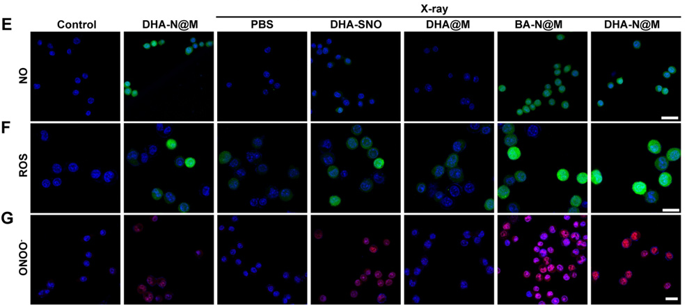
May 8th, 2025 Dr. Chen Yiting and others from the School of Pharmaceutical Sciences of Fudan University published a paper titled "Inhalable biomimetic polyunsaturated fatty acid-based." in the Journal of Nanobiotechnology (IF=10.6) The research paper on "nanoreactors for peroxynitriteaugmented ferroptosis potentiate radiotherapy in lung cancer".
The research team developed a polyunsaturated fatty acid (PUFA) -based nanorereactor DHA-N@M to act on lung tumors through inhalation administration.
This nanorreactor is composed of hados-SNO (a PUFA derivative that releases NO in response to GSH) encapsulated by macrophage cell membranes, and it has both tumor-targeting and glutathione response characteristics.
Experiments have shown that DHA-N@M significantly increases drug accumulation in lung tumors compared with intravenous injection.
Its mechanism of action is as follows: By consuming glutathione, releasing NO and generating ONOO⁻, it induces ferroptosis of tumor cells. When combined with radiotherapy, it can synergistically enhance the anti-tumor effect.
In the in situ lung cancer model, DHA-N@M combined with radiotherapy showed a tumor suppression rate of 93.91% with good safety.
This study provides a novel inhalable nanomedicine delivery system for the treatment of lung cancer. By targeting the tumor microenvironment and enhancing ferroptosis, it significantly improves the radiotherapy effect and has important potential for clinical transformation.

Here is a detailed introduction to the usage method of the glutathione detection kit for everyone.
一、Components of Kit
二、Operation steps
1. Preparation of materials and reagents
Self-prepared materials: microplate reader, distilled water, pipette and pipette tip, mortar, centrifuge, timer, wet ice, etc.
Standard: Before use, add 0.65 mL of distilled water to the tube and mix well until completely dissolved. Then, 0.05 mL of the solution was added to 0.95 mL of distilled water and mixed evenly to obtain a standard solution with a concentration of 0.5 mL /L, which was stored at 4°C.
Standard: Dye Reagent: Dissolve in 4 mL Diluent before use.
2. Sample processing
(1) Cell/Bacterial samples: Cells/bacteria (5×10⁶) were collected in centrifuge tubes. After centrifugation, the supernatant was removed, and 1mL of Assay buffer was added. After ultrasonic treatment (power 20%, ultrasonic for 3 seconds, interval 10 seconds, repeat 30 times), centrifugation was carried out at 4 ° C and 8000g for 10 minutes. The supernatant was transferred to a new centrifuge tube and stored on wet ice for testing.
(2) Tissue sample: Weigh 0.1g of the tissue, add 1mL of Assay buffer and homogenize it on wet ice. Centrifuge at 8000g at 4 ° C for 10 minutes. Transfer the supernatant to a new centrifuge tube and store it on wet ice for testing.
(3) Serum/plasma samples: Direct testing.
3. Sample loading and testing
Before loading the samples, heat all the reagents to 37℃, and then add the samples as shown in the following table.
Note:
(1)For the standard product, it can be continuously diluted by 2 times (about 6 points) to form a standard curve.
(2) For unknown samples, we suggest selecting several samples for pre-experiments to determine the sample concentration and dilution factor.
4. Standard curve
The standard curve is for reference only. A new standard curve must be made for each experiment.
Frequently Asked Questions
Q1: What if there is only one 96-Well Microplate in the kit and it is not enough for the experiment?
A1: The 96-well plates in this kit are given away for free. For subsequent experiments, regular 96-well microplate plates can be used as substitutes.
Q2: The operation manual of the reagent kit contains both the standard curve method and the formula method. Which calculation method should I choose?
A2: You can choose either of the two methods. The formula is a simple algorithm. It is recommended to calculate according to the standard curve, which will be more accurate. It should be noted that when using the standard curve method, the standard curve must be redrawn for each experiment.
Q3: If precipitation occurs during the reaction process, how should it be handled?
A3: Most precipitates or flocculent substances are caused by excessively high protein concentrations. It is recommended to aspirate the sample from the well plate, centrifuge it for a while, and then take the supernatant for testing, or add the sample to a centrifuge tube for reaction, centrifuge it, and then take the supernatant and add it to the well plate for testing.
Q4: What should I do if the instrument has no corresponding detection wavelength?
A4: If the recommended detection wavelength in the kit is not included in the instrument Settings, you can look for it near the recommended detection wavelength in the manual. Generally, there is no problem within ±10nm.
References:
[1]Chen Y, Huang X, Hu R, et al. Inhalable biomimetic polyunsaturated fatty acid-based nanoreactors for peroxynitrite-augmented ferroptosis potentiate radiotherapy in lung cancer. J Nanobiotechnology. 2025;23(1):338. Published 2025 May 8. doi:10.1186/s12951-025-03409-8